Risk Factors for Keloid Scars

If you already have a keloid, you are at increased risk of developing another. The only exception to this seems to be a single keloid on the earlobe.1 Studies suggest that the following may also be risk factors for keloids or the recurrence of a keloid after treatment:
Heredity. Analysis of patient profiles showed that the majority of people with keloids also had family members with them.1 Family history of keloids was also linked to a higher incidence of multiple keloids, and some chromosomal studies have identified genetic susceptibilities.1,4 Additionally, people born with certain genetic disorders such as Turner’s syndrome, Opitz–Kaveggia syndrome, progeria, and Rubinstein–Taybi syndrome have shown a tendency to develop keloids.12
Skin color/ethnicity. Keloids reportedly develop more often in people with darker skin, while they are never seen in people without skin pigment (albinos).5 However, some experts point out that this risk factor is only derived from decades-old observational data of spontaneous keloid development in African, Indian, Malaysian, and Caribbean native populations and not from data on post-surgical scarring.13 When keloids do occur they tend to form in areas of the body with higher amounts of skin pigment cells.5
Blood type. People with type A blood are linked to higher rates of keloid development.1
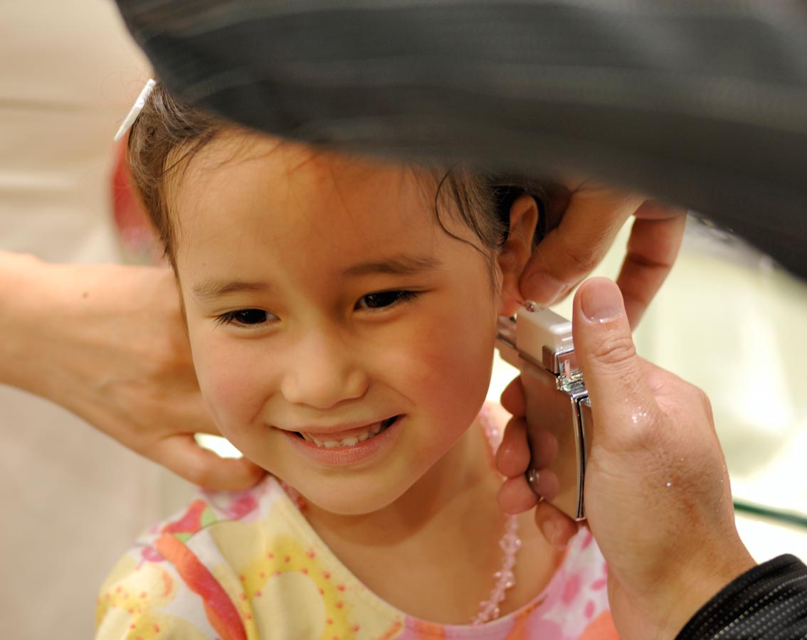
Age and gender. Younger people (ages 10-30) are more prone to keloids than older adults and the elderly. This may be due in part to changes at the molecular level in the way our body responds to wounds as we age.4 A 2005 pediatric study on occurrence of ear keloids, however, suggests that ear-piercing in children before the age of 11 may increase the risk of keloids.7 Although there appears to be a higher rate of keloids in young girls than boys, experts suggest this may be reflective of the number of girls getting their ears pierced.3
Endocrine factors. Pregnant women are more prone to keloid formation or worsening keloids, whereas post-menopausal women rarely develop keloids, while some studies suggest that after menopause keloids may shrink in size.1
Dysfunction in the thyroid and parathyroid glands (part of the endocrine system) and the hypothalamus (which interacts with the endocrine system) has also been linked to keloids, possibly because of an increased production by the glands of melanocyte-stimulating hormone (MSH). This would seem to correlate to the link between darker skin color and keloids, and MSH is normally at higher levels during pregnancy and puberty.14
Although it may seem odd that cells responsible for skin and hair color would have a role in keloid formation, melanocytes also release the neurotransmitter acetylcholine (ACh), which helps regulate how collagen-producing cells (keratinocytes) divide, grow, and stick to each other. ACh also influences inflammatory responses. All of these factors may explain why melanocytes could affect keloid formation.15
Certain systemic drugs. There may be a link between using anabolic steroids and occurrence of keloids, and keloids reportedly occurred in acne patients who have used systemic retinoids along with argon laser treatment.11
Type of wound. Burn injuries are associated with higher risk of recurrence, as is infection of any type of wound.16
Susceptible Body Areas
Certain areas of the body are more susceptible to keloid development, and race seems to have an influence on frequency of appearance:1,3
- areas with higher number of melanocytes (pigment cells)
- back
- breasts
- cheeks
- ear lobes
- legs
- stomach
- chest
- shoulder
- upper arms
Keloids on the upper extremities are more common in whites and Asians than blacks, while they appear more frequently on the lower extremities in whites and blacks than Asians.3 Whites and blacks also develop keloids in more places than Asians do.3 Keloids are rarely observed in palms of the hand and soles of the foot, where there are far fewer melanocytes.5,11 Eyelids, cornea, and mucous membranes are also less likely to experience keloid formation.1


